At a Glance
| Overview | Details |
| Who Needs Surveillance? | People with cirrhosis are an important group for whom hepatocellular carcinoma (HCC) surveillance is recommended. |
| Why Surveillance Is Needed | HCC can develop without causing noticeable symptoms, particularly in its earlier stages. |
| Main Surveillance Tests | Liver ultrasound, commonly combined with a blood test for alpha-fetoprotein (AFP). |
| How Often? | Surveillance is generally performed every 6 months in patients for whom it is indicated. |
| Does Feeling Well Mean the Risk Is Low? | No. The absence of symptoms does not reliably exclude early HCC. |
| What if Ultrasound Is Inadequate or Abnormal? | Additional imaging, such as contrast-enhanced CT or MRI, may be required depending on the findings. |
| Why Early Detection Matters | Detecting HCC at an earlier stage can increase the range of potentially curative treatment options. |
| Is Surveillance the Same for Everyone? | No. Individual risk, liver function, underlying liver disease and suitability for treatment are considered when planning surveillance. |
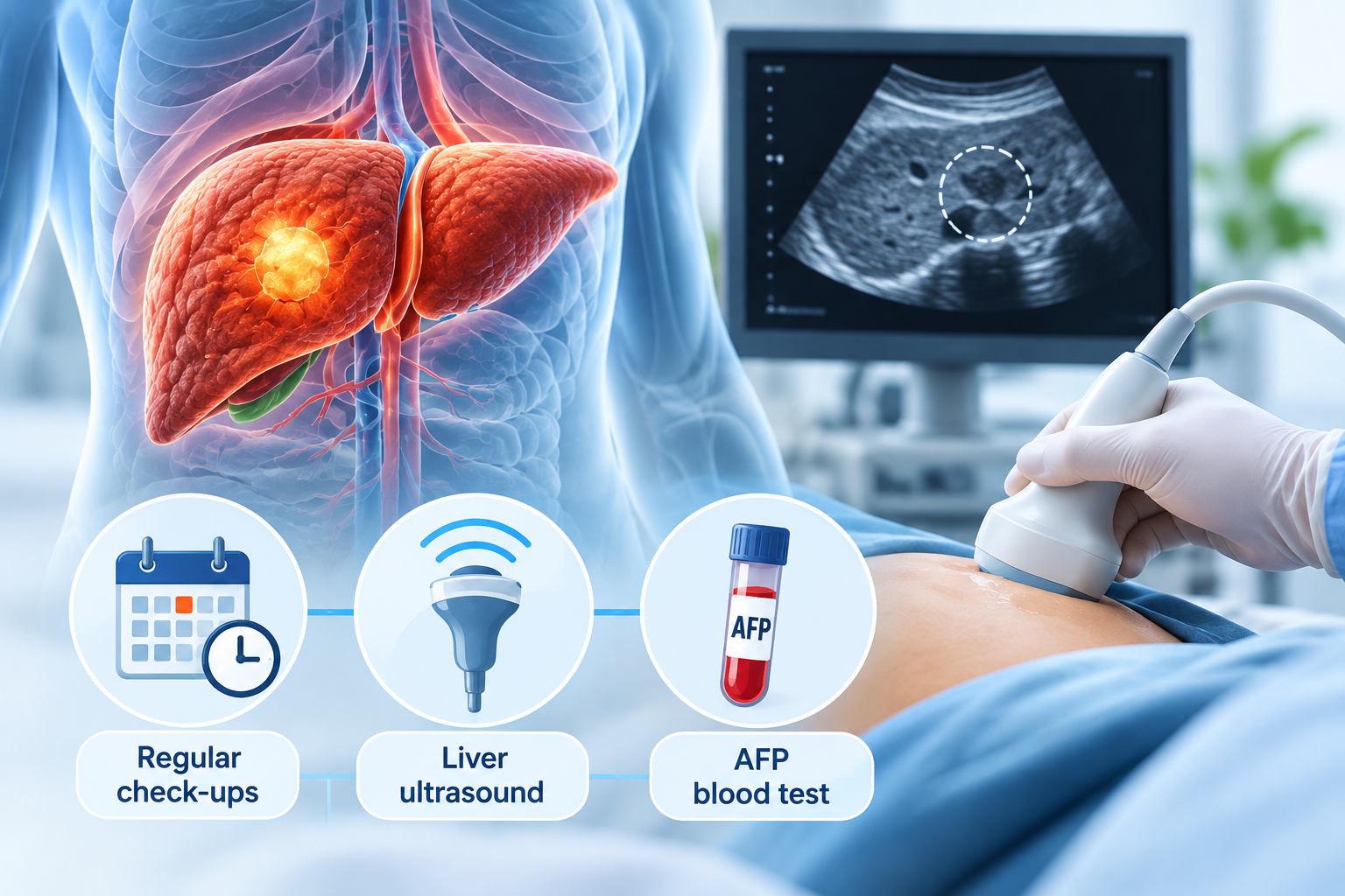
Cirrhosis does more than affect how well the liver functions. The long-term scarring and regeneration of liver tissue also increase the risk of hepatocellular carcinoma (HCC), the most common type of primary liver cancer. Importantly, this risk is not always accompanied by warning signs. A person may feel well and have no new symptoms while an early liver tumour is developing.
This is why liver cancer surveillance forms an important part of ongoing care for people with cirrhosis who meet surveillance criteria. Early HCC may cause no pain, jaundice, loss of appetite or unexplained weight loss, making symptoms an unreliable way to detect the disease at an early stage. Regular surveillance aims to identify suspicious changes before symptoms develop, when the cancer may be smaller and a wider range of treatment options may still be available.
Surveillance does not mean that everyone with cirrhosis will develop liver cancer. Rather, it provides a structured way of monitoring people at increased risk so that HCC can be detected as early as possible. In this blog, we explain why cirrhosis increases liver cancer risk, how surveillance is performed, what happens when an abnormality is detected and why regular screening remains important even when you feel well.
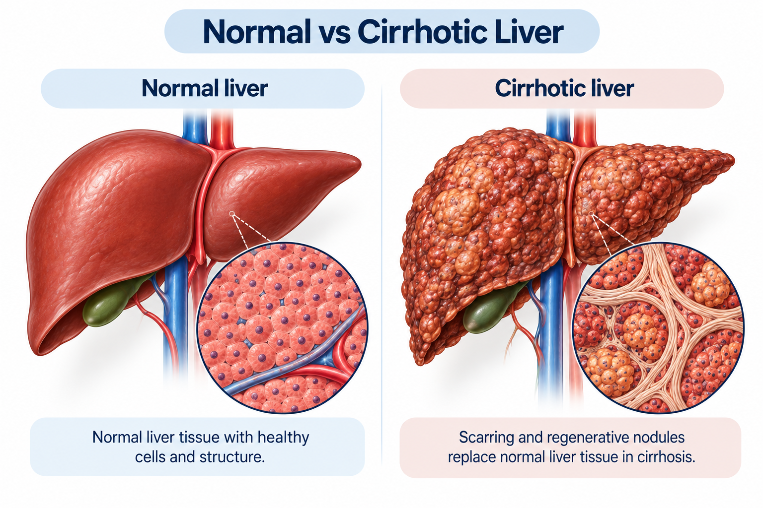
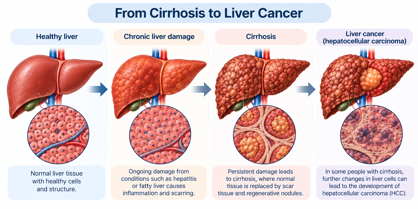
Cirrhosis is the advanced stage of scarring that can develop after prolonged liver injury. Healthy liver tissue is progressively replaced by fibrosis and regenerative nodules, which alters the normal structure of the liver. Cirrhosis can result from several underlying liver diseases, including chronic viral hepatitis, alcohol-related liver disease and metabolic liver disease.
The same long-standing processes of liver-cell injury, inflammation and regeneration that contribute to cirrhosis can also create conditions in which abnormal liver cells develop and multiply [1]. As a result, cirrhosis is one of the major risk factors for hepatocellular carcinoma. Importantly, this increased cancer risk can remain even when the underlying liver disease has been treated or brought under better control.
Having cirrhosis does not mean that liver cancer is inevitable. It means that the risk is sufficiently increased in appropriate patients for regular surveillance to become part of long-term liver care. This is fundamentally different from investigating a patient only after symptoms have developed.
Early liver cancer can develop silently because a small tumour may not initially interfere with the liver’s ability to function or affect surrounding structures. The liver has a considerable functional reserve, meaning that a substantial amount of healthy tissue can continue performing essential functions despite the presence of a developing tumour. As a result, early hepatocellular carcinoma (HCC) may produce few or no noticeable changes.
Symptoms are more likely to appear as a tumour grows, affects more liver tissue or begins to place pressure on nearby structures [2]. These may include persistent discomfort or pain in the upper-right abdomen, loss of appetite, unexplained weight loss, abdominal swelling or jaundice. However, the absence of these symptoms cannot be taken as reassurance that an early tumour is not present.
This is precisely why surveillance is important for people with cirrhosis who are at increased risk of HCC. Rather than waiting for symptoms to trigger an investigation, surveillance examines the liver at regular intervals with the aim of identifying suspicious changes while the cancer is still at an earlier stage.
Liver cancer surveillance is recommended for people whose underlying liver disease places them at a sufficiently increased risk of developing hepatocellular carcinoma (HCC) [3]. People with cirrhosis are the main group for whom regular surveillance is considered, provided their overall health and liver function are such that they could benefit from treatment if cancer were detected.
Cirrhosis can develop as a result of several chronic liver conditions, including:
However, cirrhosis is not the only situation in which surveillance may be appropriate. Some people with chronic hepatitis B have an increased risk of HCC even before cirrhosis develops, and surveillance may be recommended according to factors such as age, family history, ethnicity and other clinical risk factors [4].
The decision to begin or continue surveillance should therefore be based on an individual assessment of the underlying liver disease and HCC risk, rather than on whether a person currently has symptoms.
Liver cancer surveillance involves regularly examining the liver for early changes that may suggest hepatocellular carcinoma (HCC), even when no symptoms are present. For most people with cirrhosis who require surveillance, a liver ultrasound is performed at regular intervals, commonly together with a blood test for alpha-fetoprotein (AFP) [5].
Surveillance may include:
CT and MRI therefore have an important role in investigating abnormalities identified during surveillance, but they are not routinely required for every surveillance appointment. The imaging approach is determined by the ultrasound findings, the quality of the examination and the individual clinical situation.
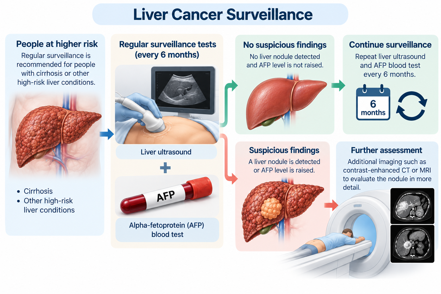
For people who require liver cancer surveillance, six months is generally considered an appropriate interval for detecting hepatocellular carcinoma (HCC) at an earlier stage [8]. This timeframe allows the liver to be reassessed regularly enough to identify a newly developing lesion or a meaningful change, without subjecting patients to unnecessarily frequent testing.
A normal surveillance result does not mean that future screening is no longer necessary. It only indicates that no concerning abnormality was identified at that particular examination. Because the underlying risk associated with cirrhosis remains, a tumour can develop between surveillance appointments even after previous ultrasound examinations have been normal.
Liver cancer surveillance should therefore be viewed as an ongoing programme, and not a one-time test. Keeping to the recommended six-month schedule reduces the likelihood of a developing tumour remaining undetected for a prolonged period and increases the opportunity for HCC to be identified while it is still at an earlier stage.
No. Alpha-fetoprotein (AFP) is useful as part of liver cancer surveillance, but it cannot reliably detect or exclude hepatocellular carcinoma (HCC) on its own. Some people with HCC have normal AFP levels, particularly when the tumour is small, while an elevated AFP can also occur in people who do not have liver cancer.
For this reason, AFP is interpreted together with liver imaging rather than as a stand-alone cancer test [9]. A rising or persistently elevated AFP level may warrant closer assessment, particularly in someone already at increased risk of HCC, but the result needs to be considered alongside ultrasound findings, previous AFP measurements and the person's underlying liver condition.
If AFP levels or imaging findings raise concern, further investigation may be recommended. This may include more detailed imaging with contrast-enhanced CT or MRI to determine whether a suspicious liver lesion is present and whether it has features consistent with HCC.
Finding a liver nodule during surveillance does not necessarily mean that liver cancer is present. Cirrhosis itself can produce regenerative and other non-cancerous nodules, so a newly detected lesion needs to be assessed according to its size, appearance and whether it has changed compared with previous scans.
Further assessment may involve:
In people at increased risk of HCC, characteristic findings on specialised contrast-enhanced imaging can sometimes establish the diagnosis without the need for a biopsy. Other nodules may remain uncertain and require additional imaging, follow-up or, in selected circumstances, tissue sampling before their nature can be determined.
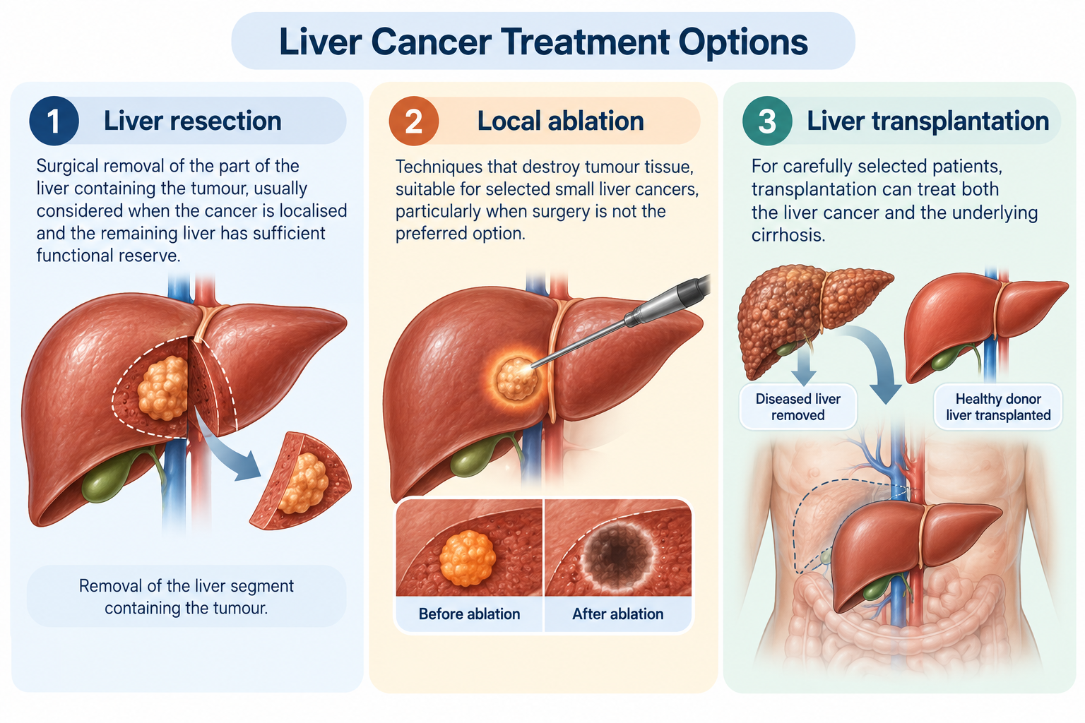
The stage at which hepatocellular carcinoma (HCC) is detected can significantly influence the treatments available. When a tumour is identified at an early stage and remains confined to the liver, some patients may be suitable for treatments with curative intent.
The choice depends not only on the size and number of tumours, but also on their location, the extent of underlying cirrhosis, liver function and the patient’s overall health. Treatment options for early-stage HCC may include:
Once HCC becomes more advanced, these potentially curative approaches may no longer be suitable, and treatment may instead involve locoregional therapies or systemic treatment.
Regular surveillance therefore aims to detect HCC while it is still at a stage when a wider range of treatment options, including potentially curative treatment, can be considered.
Not necessarily. Treating the disease responsible for cirrhosis is an important part of protecting the liver and can reduce ongoing liver damage. However, once cirrhosis has developed, successfully treating or controlling its underlying cause does not necessarily eliminate the increased risk of hepatocellular carcinoma (HCC).
For example, successful treatment of chronic hepatitis C substantially reduces the risk of HCC, but people who already have cirrhosis generally remain at increased risk even after the virus has been cleared. Similarly, stopping alcohol consumption or improving the metabolic factors contributing to liver disease can provide important benefits without necessarily removing the cancer risk associated with established cirrhosis.
Surveillance should therefore not be stopped solely because liver blood tests have improved, the underlying disease is better controlled or the person feels well. Whether ongoing HCC surveillance is required depends on the extent of liver disease and the individual’s remaining cancer risk, and should be determined by the treating doctor.
No. Liver function tests are important for monitoring cirrhosis and assessing how well the liver is functioning, but normal or stable results cannot rule out hepatocellular carcinoma (HCC). An early liver tumour may be present without causing substantial changes in routine liver enzymes or other measures of liver function.
This is because blood tests and cancer surveillance serve different purposes. Routine liver blood tests assess aspects of liver injury and function, whereas HCC surveillance is specifically intended to look for evidence of a developing liver tumour. A reassuring blood test result therefore cannot replace scheduled liver imaging.
People who have been advised to undergo HCC surveillance should continue with the recommended schedule even when their liver function tests remain stable and they have no symptoms. Neither normal blood results nor the absence of symptoms reliably excludes early HCC.
Ultrasound is commonly used for routine HCC surveillance, but it does not always provide a sufficiently clear view of the liver. In some people, factors such as body habitus or the irregular, nodular appearance of a cirrhotic liver can make small abnormalities more difficult to identify or assess.
Additional imaging may be needed when the liver cannot be adequately visualised on ultrasound or when ultrasound identifies a nodule or other abnormality that requires further characterisation. Depending on the findings, this may involve contrast-enhanced multiphasic CT or MRI, which can provide more detailed information about the lesion and its pattern of contrast enhancement.
This does not mean that every person undergoing HCC surveillance routinely requires CT or MRI. More detailed imaging is generally used when the quality of ultrasound is inadequate or when a finding needs further investigation, with the appropriate imaging approach determined according to the individual clinical situation.
Regular surveillance should not delay medical assessment if new symptoms develop between scheduled appointments. A previous normal ultrasound only reflects the findings at the time it was performed, and people with cirrhosis should seek medical advice if they develop a significant or unexplained change in their health.
Symptoms that warrant medical assessment may include:
These symptoms are not specific to liver cancer and can occur with cirrhosis itself or other medical conditions. However, they should be assessed when they develop rather than attributed to existing liver disease or left until the next scheduled surveillance appointment.
Feeling well can make repeated surveillance appointments seem unnecessary, particularly when previous scans have been normal and blood tests remain stable. However, the absence of symptoms is precisely why surveillance is needed: early HCC can develop without causing noticeable changes in how a person feels.
Each surveillance examination assesses the liver at a particular point in time. A normal result six months earlier does not mean that a new lesion cannot subsequently develop, nor does stable liver function eliminate the cancer risk associated with cirrhosis.
Keeping to the recommended surveillance schedule therefore provides repeated opportunities to identify HCC before symptoms develop. If surveillance appointments have been missed or there has been a prolonged gap since the last examination, the appropriate timing of the next assessment should be discussed with the treating doctor.
Cirrhosis is an important risk factor for hepatocellular carcinoma (HCC), and the disease can develop at an early stage without causing noticeable symptoms or significant changes in routine liver blood tests. Liver cancer surveillance is therefore based on a person’s underlying risk rather than on the presence of symptoms, allowing suspicious changes to be identified before they would otherwise become clinically apparent.
For people with cirrhosis who meet surveillance criteria, regular liver ultrasound, commonly accompanied by an alpha-fetoprotein (AFP) blood test, provides an opportunity to detect HCC at an earlier stage. Surveillance generally continues at approximately six-month intervals because a normal examination reflects the liver at that point in time and does not eliminate the possibility of HCC developing subsequently.
When ultrasound identifies an abnormality or cannot adequately assess the liver, further investigation with contrast-enhanced CT or MRI may be required. If you have cirrhosis or another chronic liver condition, schedule a consultation with a hepatobiliary specialist at Tan Siong San Surgery to assess your liver cancer risk and determine an appropriate surveillance plan.


Tan Siong San Surgery. © All Rights Reserved 2025


