BOOK AN APPOINTMENT
BOOK AN APPOINTMENT

Pancreatic Cyst Found by Accident on a Scan — Should You Be Worried?

October 23, 2026
OverviewDetails
ConditionPancreatic cysts
How are they discoveredOften incidentally during an ultrasound, CT scan or MRI performed for another condition
Are they cancerous?Many are benign, but certain cyst types can develop into cancer
What determines the risk?Cyst type, size, imaging characteristics, growth, symptoms and other clinical findings
Further assessmentMRI with magnetic resonance cholangiopancreatography (MRCP), CT or endoscopic ultrasound (EUS), depending on the findings
Is regular surveillance necessary?Some cysts require periodic imaging, while others may not need routine follow-up
When treatment may be consideredWhen the cyst has concerning features, causes significant symptoms or is associated with suspected or confirmed malignancy
Pancreatic Cyst Singapore
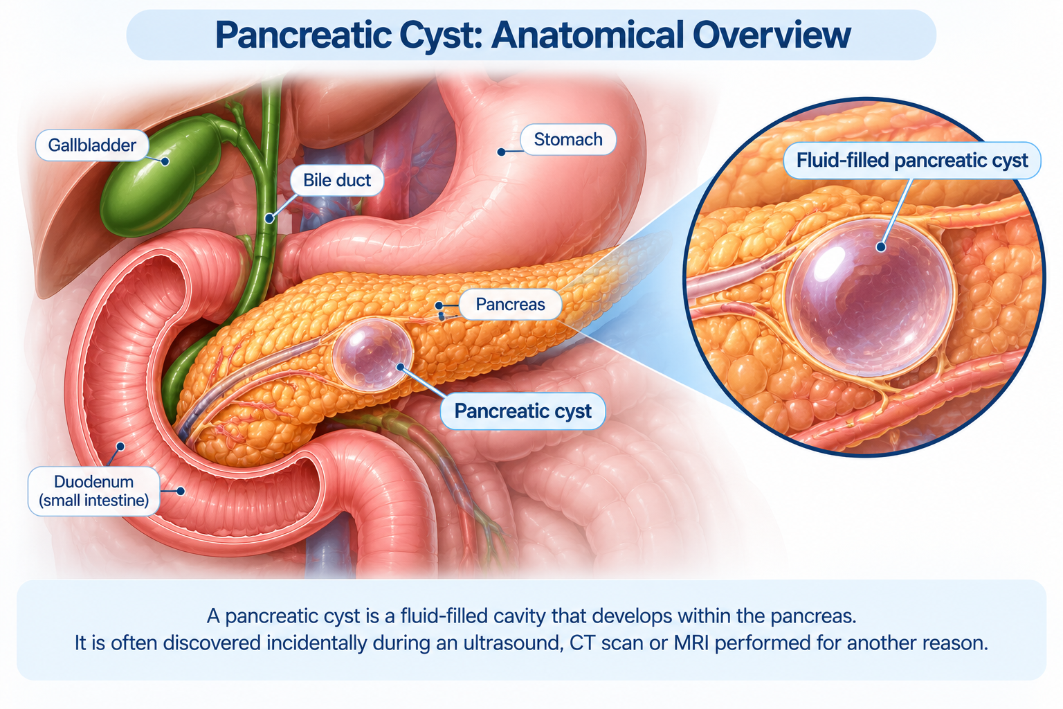
A pancreatic cyst is a fluid-filled lesion within the pancreas that is often discovered incidentally during an ultrasound, CT scan or MRI performed for another medical reason.

An unexpected finding of a pancreatic cyst during an abdominal scan can raise immediate concerns, particularly because of the association between pancreatic disease and cancer. Yet pancreatic cysts are not all alike. Many are discovered in people who have no pancreatic symptoms, and some are benign lesions that may never cause problems. Others, however, have the potential to develop into pancreatic cancer. 

The difficulty is that a pancreatic cyst cannot be assessed by its presence or size alone. Its internal structure, relationship with the pancreatic duct, changes over time and any associated symptoms can provide important clues about its nature. A small cyst may require further investigation if it has suspicious features, while another cyst may be suitable for surveillance without immediate intervention. 

Understanding the type of pancreatic cyst is therefore the starting point for deciding what happens next. In this blog, we explain why pancreatic cysts are found incidentally, how benign and potentially precancerous cysts differ, which findings warrant closer attention and when surveillance, further investigation or specialist treatment may be appropriate.

What Exactly is a Pancreatic Cyst?

A pancreatic cyst is a fluid-containing lesion that develops within or around the pancreas, an organ located behind the stomach that produces digestive enzymes and hormones involved in regulating blood sugar. Pancreatic cysts encompass several different conditions, ranging from benign fluid collections to cystic tumours with varying degrees of malignant potential.

Many pancreatic cysts are detected incidentally during CT or MRI examinations performed for unrelated abdominal symptoms or other medical conditions. Improved imaging technology has made it increasingly common to identify small cysts that might otherwise have remained undetected.

Not every pancreatic cyst requires treatment, but identifying its type is important because the approach to management differs considerably. Some cysts have characteristic imaging features that support a benign diagnosis, while others require closer examination of their internal structure and relationship with the pancreatic duct.

Why are Pancreatic Cysts Often Found Incidentally?

Many pancreatic cysts develop without causing pain, digestive difficulties or other noticeable symptoms. Small cysts may not interfere with the pancreatic duct or surrounding structures, allowing them to remain undetected for years.

They are frequently discovered during imaging undertaken for another reason, such as investigating abdominal discomfort, examining the liver or gallbladder, or assessing an unrelated medical condition. The finding may appear in a radiology report even when there was no previous suspicion of pancreatic disease.

The increased detection of incidental pancreatic cysts does not mean that all such lesions are dangerous. It does, however, create an important clinical question: does the cyst have features consistent with a benign lesion, or does it belong to a category that requires surveillance or further investigation?

The answer depends on the cyst’s characteristics and the patient’s wider clinical circumstances, rather than simply on the fact that it was discovered unexpectedly.

What are the Different Types of Pancreatic Cysts?

Pancreatic cysts are not a single disease. Different types have distinct causes, imaging appearances and implications for treatment or follow-up. Establishing which type is present helps determine whether the cyst can be left alone, requires ongoing surveillance or needs further assessment.

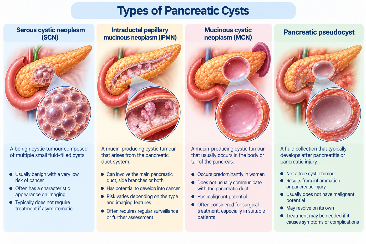
  • Serous cystic neoplasms (SCNs) — These are generally benign pancreatic cystic tumours with a very low risk of malignant transformation [1]. They often contain multiple small fluid-filled spaces and may have a characteristic appearance on imaging. When the diagnosis is confident and the lesion is not causing symptoms, surgery is usually unnecessary.
  • Intraductal papillary mucinous neoplasms (IPMNs) — These are mucin-producing cystic tumours arising from the pancreatic duct system [2]. They may involve the main pancreatic duct, its side branches or both. IPMNs are important because some can progress to high-grade dysplasia or invasive cancer. Their risk varies according to the type of IPMN and the presence of concerning imaging or clinical features.
  • Mucinous cystic neoplasms (MCNs) — These are mucin-producing cystic tumours that occur predominantly in women and are usually located in the body or tail of the pancreas [3]. Unlike IPMNs, they generally do not communicate with the pancreatic duct. MCNs have malignant potential, although not every lesion contains or will develop cancer.
  • Pancreatic pseudocysts — These are fluid collections that typically develop following acute pancreatitis, chronic pancreatitis or pancreatic injury [4]. They are not true cystic neoplasms and do not have the same precancerous potential as mucinous cystic tumours. Their management depends on factors such as symptoms, infection, obstruction and other complications.
  • Other pancreatic cystic lesions — Less common conditions include solid pseudopapillary neoplasms, cystic neuroendocrine tumours and other unusual pancreatic lesions. Some require specialist investigation because their behaviour and treatment differ from those of more common pancreatic cysts.

These distinctions are particularly important because the term “pancreatic cyst” does not indicate whether a lesion is benign, precancerous or malignant. Further imaging or specialist assessment may be needed when its nature cannot be established confidently from the initial scan.
Types of Pancreatic Cysts Singapore
The four main types of pancreatic cysts include serous cystic neoplasms, intraductal papillary mucinous neoplasms, mucinous cystic neoplasms and pancreatic pseudocysts, each with different characteristics, cancer risks and management considerations.

Can an Incidental Pancreatic Cyst Be Cancerous?

Yes, certain pancreatic cysts can contain cancer or develop malignant changes over time, but this does not mean that an incidental cyst should be assumed to be cancerous. Many pancreatic cysts are benign, and even among cyst types with malignant potential, progression to cancer is not inevitable. 

The principal concern is identifying the smaller group of lesions that may already contain advanced abnormal cells or have characteristics associated with a higher risk of progression. Mucin-producing cysts, particularly IPMNs and MCNs, receive closer attention because of their potential to develop into cancer [5]. A small branch-duct IPMN without concerning features may be managed through imaging surveillance, whereas a lesion with an enhancing solid component or significant pancreatic duct dilatation may require more urgent specialist evaluation.

The aim of assessment is therefore to identify potentially dangerous cysts without exposing patients with low-risk lesions to unnecessary pancreatic surgery. This distinction is especially important because pancreatic surgery is a major procedure with potential complications, and its benefits must be weighed against the estimated risk associated with the cyst. 

Which Pancreatic Cyst Features Require Closer Attention?

The imaging characteristics of a pancreatic cyst are central to assessing its potential risk. Certain findings can suggest a greater likelihood of advanced cellular changes or malignancy, particularly in mucin-producing cysts. These features are assessed alongside the patient's symptoms, medical history and changes observed on previous scans.

Features that may warrant further investigation include:

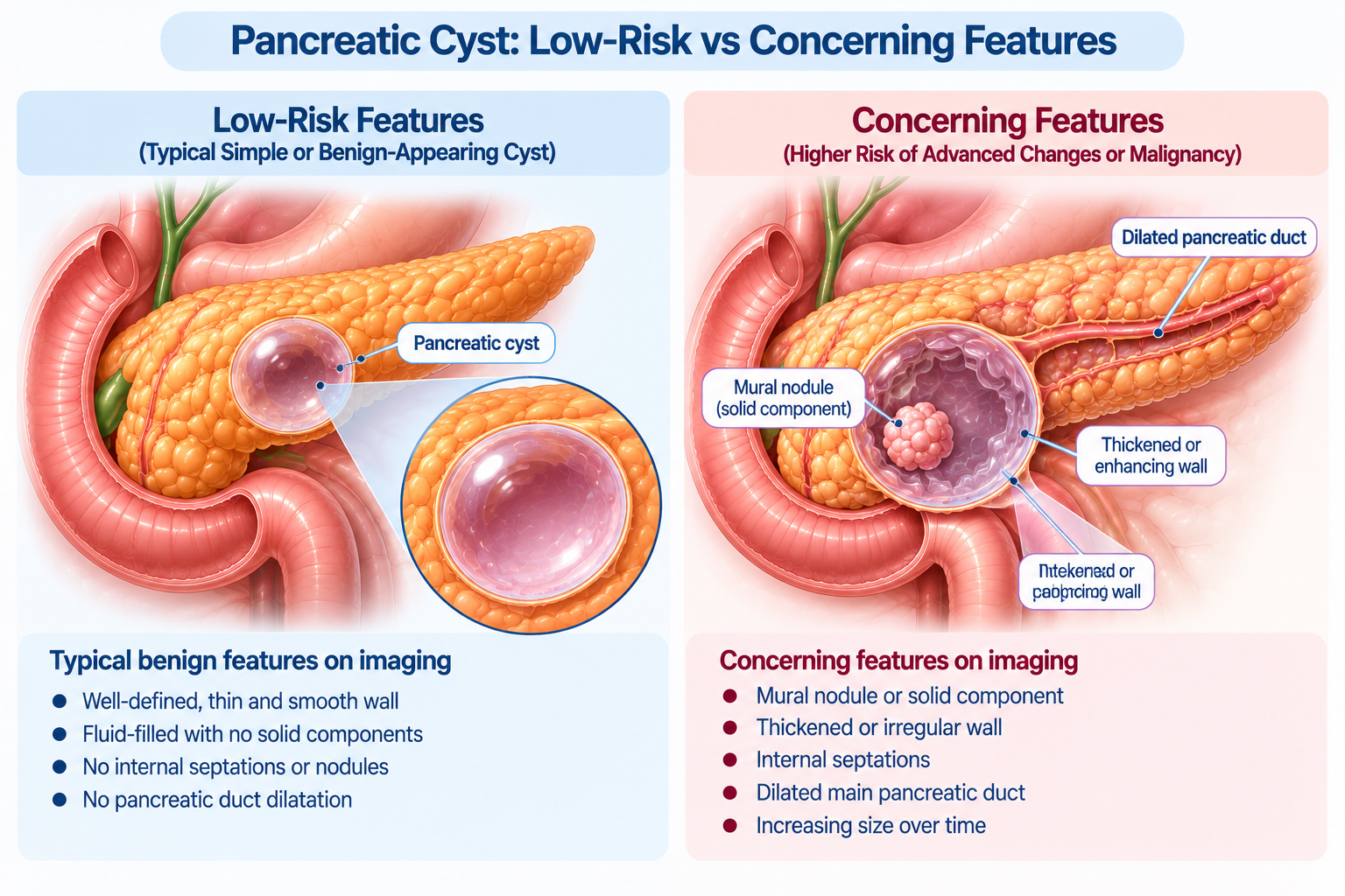
  • A solid component or mural nodule — A growth projecting from the cyst wall, particularly one that enhances after contrast administration, can be an important warning feature.
  • Dilatation of the main pancreatic duct — Enlargement of the duct may indicate involvement of the duct system and can increase concern in certain cyst types.
  • Thickened or enhancing cyst walls — These may require closer evaluation, particularly when accompanied by other atypical findings.
  • Increasing cyst size or significant growth — Larger cysts and those growing relatively quickly may warrant additional assessment, although growth alone does not establish malignancy.
  • Jaundice associated with a pancreatic head cyst — Yellowing of the skin or eyes caused by obstruction of the bile duct is a particularly concerning finding.
  • Pancreatitis associated with the cyst — An episode of pancreatic inflammation potentially caused by the lesion may influence the need for further investigation.
  • New-onset or worsening diabetes — In the context of a pancreatic cyst, an unexplained change in blood sugar control may be relevant to risk assessment.
  • Elevated carbohydrate antigen 19-9 (CA 19-9) — This blood marker may provide additional information, although it cannot diagnose pancreatic cancer on its own.

These findings do not all carry the same level of risk. The 2024 International Evidence-Based Kyoto Guidelines distinguish between worrisome features, which generally prompt closer investigation, and high-risk stigmata, which may indicate a stronger need to consider surgery. Their interpretation is particularly relevant to IPMNs and should not be applied indiscriminately to every type of pancreatic cyst [6]. 
Pancreatic Cyst Risk Features Singapore
Pancreatic cysts with smooth, thin walls and no solid components may have lower-risk features, while mural nodules, wall thickening or pancreatic duct dilatation may warrant further investigation.

Does the Size of a Pancreatic Cyst Determine Whether It is Dangerous?

Cyst size is an important part of pancreatic cyst assessment, but it cannot establish whether a lesion is benign or malignant on its own. A relatively large cyst with characteristic features of a serous cystic neoplasm may be benign, while a smaller mucin-producing cyst with an enhancing mural nodule may require more detailed investigation.

Rather than relying on a single measurement, doctors consider several questions:

  • What type of pancreatic cyst is most likely?
  • Does the cyst contain a mural nodule or solid component?
  • Is the main pancreatic duct enlarged?
  • Has the cyst increased in size since previous imaging?
  • Are there symptoms such as jaundice, pancreatitis or unexplained weight loss?
  • Does the cyst have other features associated with a higher risk of malignancy?

For IPMNs, a cyst measuring 3 cm or more is generally considered a worrisome feature under the 2024 Kyoto guidelines, but this threshold is not, by itself, an indication for surgery [7]. Other findings, the patient’s overall health and the potential benefits and risks of intervention remain essential considerations.

The significance of cyst size therefore depends on the type of lesion and the other findings present. A larger measurement may justify closer assessment without necessarily making surgical removal the appropriate next step. 

Can a Pancreatic Cyst Cause Symptoms?

Many pancreatic cysts cause no symptoms and are discovered only because imaging was performed for another reason. Symptoms are more likely when a cyst becomes sufficiently large to affect nearby structures, obstructs pancreatic or bile drainage or is associated with inflammation or another complication.

Possible symptoms include:

  • Persistent or recurrent upper abdominal pain
  • Pain extending towards the back
  • Nausea or vomiting
  • Feeling full after eating relatively small amounts
  • Jaundice, particularly if bile drainage is obstructed
  • Episodes of pancreatitis
  • Unexplained weight loss or loss of appetite

These symptoms are not specific to pancreatic cysts. Gallbladder disorders, stomach conditions, pancreatitis and other abdominal diseases can produce similar complaints. The presence of a pancreatic cyst on imaging therefore does not establish that it is responsible for a patient’s symptoms.

When symptoms are present, further assessment may be needed to determine whether the cyst’s size, location and imaging features provide a plausible explanation or whether another condition should be investigated.

How are Pancreatic Cysts Diagnosed and Assessed?

Pancreatic cysts are commonly identified on CT or MRI performed for another medical reason. Once a cyst is detected, the next step is to establish its likely type, assess features associated with malignancy and determine whether additional investigation or surveillance is appropriate.

Assessment may involve:

  • MRI with magnetic resonance cholangiopancreatography (MRCP) — MRI provides detailed images of the pancreas and cyst contents, while MRCP helps assess the pancreatic duct system and whether a cyst communicates with it. MRI/MRCP is often preferred for characterising and monitoring pancreatic cysts because it avoids ionising radiation.
  • Contrast-enhanced CT — CT can help assess the cyst, surrounding pancreatic tissue and associated abnormalities. It may be particularly useful when further anatomical detail is required or when a suspicious lesion needs more extensive evaluation.
  • Endoscopic ultrasound (EUS) — This examination involves passing a flexible endoscope through the mouth into the stomach and duodenum. An ultrasound probe at its tip provides detailed images of the pancreas from close range, allowing smaller structures, mural nodules and other features to be assessed more closely.
  • EUS-guided fluid sampling — In selected cases, fluid may be collected from the cyst using a fine needle under endoscopic ultrasound guidance. The sample can be assessed for biochemical markers, cytology or molecular findings that may help clarify the cyst type or identify concerning changes. Sampling is not necessary for every cyst and is generally considered when the results could influence management.
  • Blood tests and clinical assessment — Relevant investigations may include blood glucose measurements, liver blood tests or CA 19-9 in appropriate circumstances. These findings support the overall assessment but cannot reliably determine whether a cyst is cancerous in isolation.

A clearly characterised, low-risk cyst may not require all these investigations. More detailed assessment is generally reserved for lesions whose nature remains uncertain or whose imaging or clinical features raise concern. 

When Does a Pancreatic Cyst Need a Specialist’s Opinion?

Not every pancreatic cyst requires surgery or extensive investigation. However, specialist assessment becomes important when the initial imaging does not establish the cyst type, when concerning features are present or when a decision is needed about surveillance or treatment.

A specialist opinion may be appropriate when:

  • The cyst contains a mural nodule, solid component or other suspicious imaging features
  • The main pancreatic duct is dilated
  • The cyst has increased significantly in size
  • The diagnosis remains uncertain after initial imaging
  • The patient develops jaundice or pancreatitis potentially related to the cyst
  • There is unexplained weight loss or new-onset diabetes alongside the cyst
  • A mucinous cystic neoplasm or IPMN with concerning features is suspected
  • The cyst may require ongoing surveillance or surgical treatment

The purpose of specialist assessment is not necessarily to recommend removal. In many cases, it is to establish the likely diagnosis, determine the level of risk and decide whether observation, further imaging, endoscopic assessment or treatment is appropriate.

A hepatopancreatobiliary (HPB) specialist can review the imaging findings alongside the patient’s medical history and overall health, particularly when the potential benefits of intervention need to be weighed against the risks of pancreatic surgery.

Do Pancreatic Cysts Require Regular Follow-Up Scans?

Some pancreatic cysts require surveillance because their appearance may change over time or because they belong to a category with malignant potential. However, routine follow-up is not necessary for every pancreatic cyst, and the appropriate schedule depends on the type of lesion, its characteristics and the patient's circumstances.

For example, a confidently diagnosed asymptomatic serous cystic neoplasm may not require long-term routine imaging. In contrast, many branch-duct IPMNs without high-risk features are monitored using MRI or other appropriate imaging to identify changes that could influence management.

The surveillance plan may take into account:

  • Cyst type — Mucin-producing cysts generally require different consideration from clearly benign lesions.
  • Cyst size — Size can influence surveillance intervals, particularly for branch-duct IPMNs.
  • Imaging characteristics — Nodules, pancreatic duct changes or other concerning findings may justify closer monitoring.
  • Changes over time — Growth or the development of new features may lead to additional investigation.
  • Age and overall health — Surveillance is most useful when detecting a concerning change could lead to treatment that the patient is fit and willing to undergo.
  • Previous investigations — Earlier scans and specialist findings can help establish an appropriate monitoring schedule.

There is no single surveillance interval suitable for every pancreatic cyst. Published guidelines also differ in their recommendations about how frequently certain cysts should be monitored and when surveillance may be discontinued.

Patients should therefore follow an individualised plan rather than assume that every incidental pancreatic cyst requires annual imaging or that surveillance can safely stop after a fixed number of normal examinations. 

What Happens if a Pancreatic Cyst Changes During Surveillance?

Changes detected during surveillance do not necessarily mean that a cyst has become cancerous. Some pancreatic cysts grow slowly over time without developing malignant features. However, a significant change in size, the appearance of a new mural nodule or increasing pancreatic duct dilatation may alter the estimated risk and require further assessment.

When a change is identified, the treating specialist may recommend a shorter interval before the next scan, more detailed imaging or endoscopic ultrasound. In selected cases, EUS-guided sampling may help clarify whether the cyst has developed concerning characteristics.

The decision depends on the nature of the change rather than growth alone. A small increase in size without other suspicious findings may be managed differently from a cyst that develops an enhancing solid component or causes obstructive jaundice.

Surveillance is therefore an active process of reassessment. Its purpose is to identify meaningful changes that could influence treatment decisions while avoiding unnecessary intervention for cysts that remain low risk.

When is Surgery Considered for a Pancreatic Cyst?

Most incidentally detected pancreatic cysts do not require immediate surgery. Surgical treatment is generally considered when a lesion has features associated with a substantial risk of malignancy, when cancer is suspected or confirmed, or when certain cysts cause significant symptoms or complications.

The decision depends on several factors, including the likely cyst type, the presence of high-risk findings, its location within the pancreas and the patient's suitability for surgery.

Possible procedures include:

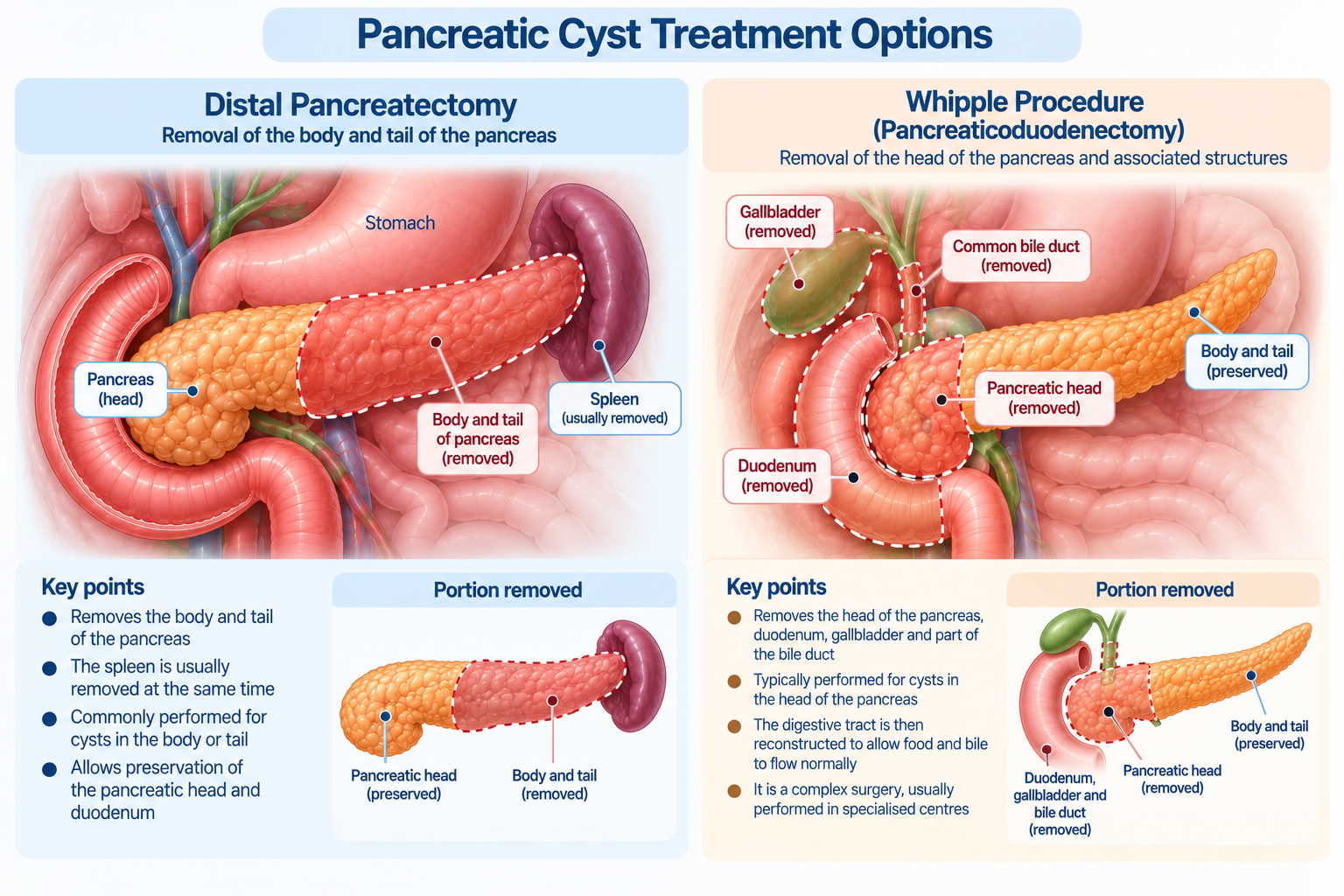
  • Distal pancreatectomy — This involves removing the body or tail of the pancreas and may be considered for selected cystic neoplasms located in these regions. Depending on the condition, the spleen may also need to be removed.
  • Pancreaticoduodenectomy (Whipple procedure) — This is a major operation that may be necessary for selected concerning or malignant lesions involving the head of the pancreas. It involves removing the pancreatic head and other associated structures, followed by surgical reconstruction.
  • Other pancreatic resections — In carefully selected circumstances, a different procedure may be appropriate depending on the location and nature of the lesion.

Pancreatic surgery carries important risks, including pancreatic leakage, infection, bleeding and potential long-term effects on digestion or blood sugar regulation. These considerations are particularly relevant when the cyst has a low estimated risk of malignant progression.

For this reason, surgery is not recommended solely because a pancreatic cyst has been detected. The decision should follow an assessment of the likely benefits of removing the lesion against the risks of the proposed operation, ideally involving an experienced pancreatic multidisciplinary team. 
Pancreatic Cyst Surgical Treatments Singapore
Surgical treatment for selected pancreatic cysts may involve distal pancreatectomy, which removes the body and tail of the pancreas, or the Whipple procedure, which removes the pancreatic head and associated structures.

Key Takeaway

An incidental pancreatic cyst is not necessarily a sign of cancer, and many lesions can be managed without surgery. However, pancreatic cysts differ substantially in their behaviour. While some are benign, others, particularly certain mucin-producing cysts, have the potential to develop malignant changes. 

Management depends on more than cyst size. Features such as mural nodules, pancreatic duct dilatation, significant growth, jaundice or other concerning clinical findings may warrant additional assessment. Some cysts require regular imaging surveillance, while others may need little or no follow-up once their benign nature has been confidently established. Surgery is generally reserved for selected lesions where the expected benefits justify the risks of pancreatic intervention.

If a pancreatic cyst has been identified on your scan, schedule a consultation with a specialist at Tan Siong San Surgery to review the findings and determine whether further investigation, surveillance or treatment is appropriate.

Frequently Asked Questions (FAQs)

Does a pancreatic cyst mean I have pancreatic cancer?

No. Many pancreatic cysts are benign, and even cyst types with malignant potential do not necessarily progress to cancer. The risk depends on the type of cyst and whether it has concerning clinical or imaging features.

Can a pancreatic cyst turn into cancer?

Certain cystic neoplasms, particularly IPMNs and MCNs, have the potential to develop malignant changes. Other types, such as serous cystic neoplasms, are generally benign. Identifying the cyst type is important when estimating cancer risk.

Can a small pancreatic cyst be dangerous?

Yes, although many small pancreatic cysts are low risk. A small lesion with an enhancing mural nodule or significant pancreatic duct abnormalities may require closer assessment. Size should always be interpreted alongside other findings.

Is a 2 cm pancreatic cyst considered dangerous?

A 2 cm measurement alone cannot establish whether a pancreatic cyst is dangerous. The likely cyst type, internal features, pancreatic duct findings, symptoms and any changes on previous imaging are important in determining whether surveillance or further investigation is appropriate.

Does every pancreatic cyst need an MRI?

No. MRI/MRCP is frequently preferred for further characterisation and surveillance, but it is not necessary in every case. The need for additional imaging depends on the information available from the initial scan and whether the result could influence management.

Is endoscopic ultrasound necessary for every pancreatic cyst?

No. Endoscopic ultrasound is generally considered when more detailed assessment is required, particularly if the cyst has concerning features or its nature remains uncertain. Many low-risk cysts can be assessed and monitored using cross-sectional imaging alone.

Can pancreatic cysts disappear on their own?

Some inflammatory fluid collections, including pancreatic pseudocysts, may resolve as the underlying pancreatitis improves. True pancreatic cystic neoplasms generally do not disappear spontaneously, although many remain stable and may not require treatment.

Can a pancreatic cyst cause pancreatitis?

Yes. Certain pancreatic cysts, particularly IPMNs, may interfere with pancreatic duct drainage and contribute to pancreatitis. However, pancreatitis has several other possible causes, so the relationship between a cyst and an episode of inflammation needs to be assessed.

Does a growing pancreatic cyst always require surgery?

No. Growth can be a reason for closer evaluation, but it does not independently establish malignancy or make surgery necessary. The rate of growth and any accompanying suspicious features are important considerations.

What type of specialist should assess a pancreatic cyst?

A hepatopancreatobiliary (HPB) specialist or a gastroenterologist with expertise in pancreatic cysts can assess the lesion and recommend appropriate investigations. Complex or high-risk lesions may benefit from review by a multidisciplinary pancreatic team.

References

  1. Hu, F., Hu, Y., Wang, D., Ma, X., Yue, Y., Tang, W., Liu, W., Wu, P., Peng, W., & Tong, T. (2022). Cystic neoplasms of the pancreas: Differential diagnosis and radiology correlation. Frontiers in Oncology, 12, 860740. https://doi.org/10.3389/fonc.2022.860740 
  2. Paramythiotis, D., Karlafti, E., Fotiadou, G., Charalampidou, M., Karakatsanis, A., Ioannidis, A., & Michalopoulos, A. (2023). Pancreatic intraductal papillary mucinous neoplasms: A narrative review. Acta Medica Lituanica, 30(1), 53–65. https://doi.org/10.15388/Amed.2023.30.1.6 
  3. Ikuta, S., Aihara, T., Yasui, C., Iida, H., Yanagi, H., Mitsunobu, M., Kakuno, A., & Yamanaka, N. (2008). Large mucinous cystic neoplasm of the pancreas associated with pregnancy. World Journal of Gastroenterology : WJG, 14(47), 7252–7255. https://doi.org/10.3748/wjg.14.7252 
  4. Pan, G., Wan, M. H., Xie, K.-L., Li, W., Hu, W.-M., Liu, X.-B., Tang, W.-F., & Wu, H. (2015). Classification and management of pancreatic pseudocysts. Medicine, 94(24), e960. https://doi.org/10.1097/MD.0000000000000960 
  5. Kruse, D. E., & Paulson, E. K. (2024). The incidental pancreatic cyst: When to worry about cancer. Korean Journal of Radiology, 25(6), 559–564. https://doi.org/10.3348/kjr.2024.0085 
  6. International evidence-based Kyoto guidelines for the management of intraductal papillary mucinous neoplasm of the pancreas. https://pubmed.ncbi.nlm.nih.gov/38182527/ 
  7. Danti, G., Scalzone, L., Mattolini, L., Anichini, M., Treballi, F., Calistri, L., Cozzi, D., & Miele, V. (2026). Pancreatic cystic lesions: From basic knowledge to recent guidelines. Journal of Clinical Medicine, 15(2), 585. https://doi.org/10.3390/jcm15020585 

Dr. Tan Siong San

Adjunct Assistant Prof (Duke-NUS)
MBBS (NUS)
MRCS (Edinburgh)
FRCS (Edinburgh)
M.Med (Singapore)
FAMS

Committed to Giving My Best for Every Patient.

This article has been medically reviewed by Dr. Tan Siong San
Dr Tan is a Senior Consultant Surgeon with over 25 years of clinical experience in the field of Hepato-Pancreato-Biliary (HPB) surgery treating conditions affecting gallbladder, liver and pancreas.

"Personalised specialist care, tailored to the unique needs of every patient"

Get an Appointment!

Book a consultation with Dr. Tan Siong San at Mount Elizabeth Novena for personalised, timely surgical care in a comfortable and central location.


    LOCATION
    38 Irrawaddy Road, #09-58 Mount Elizabeth Novena Specialist Centre, Singapore 329563.
    OPENING HOURS
    Monday to Friday

    8.30am to 12.30pm,
    1.30pm to 5.30pm

    Saturday

    8.30am to 12.30pm

    Sunday & Public Holidays
    Closed

    Tan Siong San Surgery. © All Rights Reserved 2025

    cross