At a glance
| Overview | Details |
| Conditions Covered | Gallstones and gallbladder polyps |
| Main Difference | Gallstones are solid deposits that form within bile, while gallbladder polyps are growths arising from the gallbladder wall |
| How They Are Found | Both are commonly detected on abdominal ultrasound, sometimes incidentally |
| Symptoms | Gallstones are more likely to cause biliary pain and complications; gallbladder polyps are usually asymptomatic |
| Main Health Concern | Gallstones may cause inflammation, bile duct blockage or pancreatitis, while gallbladder polyps are assessed mainly for features associated with malignancy risk |
| Role of Size | Stone size alone usually does not determine treatment; polyp size is an important factor in deciding whether surveillance or surgery is needed |
| Assessment | Ultrasound is usually the first-line investigation, with attention to mobility, acoustic shadowing, size, morphology and gallbladder wall features |
| Treatment Approach | Asymptomatic gallstones often require no treatment, while symptomatic or complicated stones may require surgery; polyps may be observed, monitored or removed depending on size and risk features |
| Surgery | Cholecystectomy removes the entire gallbladder and may be recommended for symptomatic gallstones or gallbladder polyps that meet criteria for surgical management |
| When to Seek Medical Advice | Severe or persistent upper abdominal pain, fever, jaundice, persistent vomiting or worsening symptoms should be medically assessed |
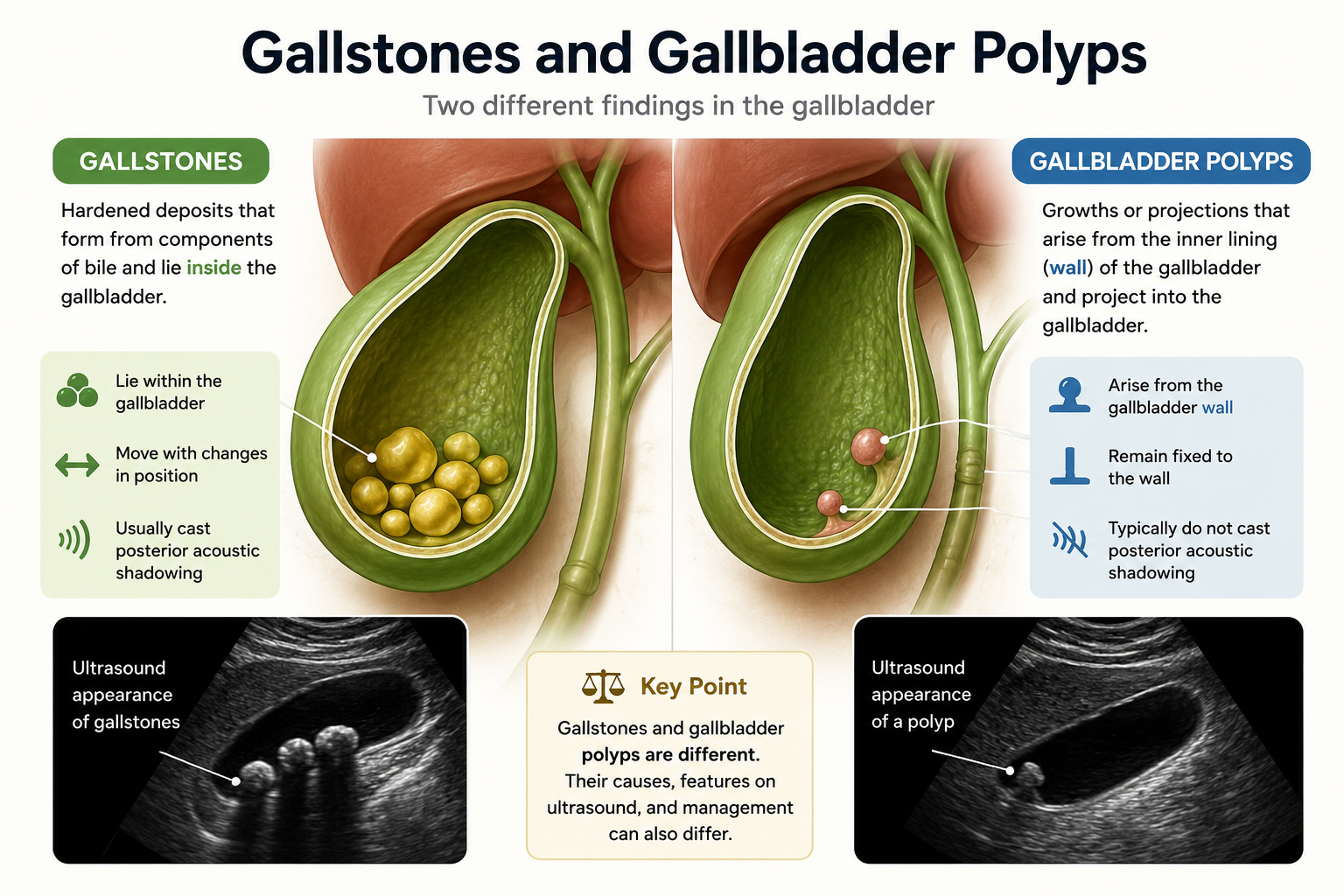
An abdominal ultrasound may reveal an unexpected abnormality in the gallbladder, even when the scan was performed for an entirely different reason. When the report identifies a gallstone or gallbladder polyp, it is important to understand that these findings arise differently and are assessed for different reasons. Their presence alone does not necessarily indicate a serious condition or mean that treatment is required.
Gallstones are solid deposits that develop from components of bile within the gallbladder, whereas gallbladder polyps are projections arising from its inner lining. Many gallstones remain completely asymptomatic, and most gallbladder polyps detected on ultrasound are benign. However, gallstones can become clinically important when they cause pain, obstruction or inflammation, while the significance of a polyp depends largely on characteristics such as its size, appearance, growth and associated risk factors.
In this blog, we explain the differences between gallstones and gallbladder polyps, the symptoms and complications they may cause, how they are assessed on imaging and the factors that determine whether monitoring or treatment may be needed.
The gallbladder is a small organ located beneath the liver. It stores and concentrates bile, a digestive fluid produced by the liver that helps with the digestion and absorption of fats. Gallstones and gallbladder polyps can both be identified during an ultrasound examination of this organ, but they originate in different places.
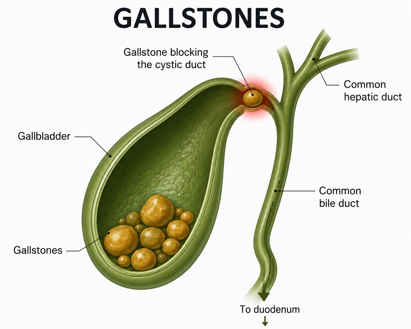
A gallstone develops within bile when certain substances crystallise and gradually form solid material [1]. The resulting stone lies within the cavity of the gallbladder and is usually able to move when the patient’s position changes.
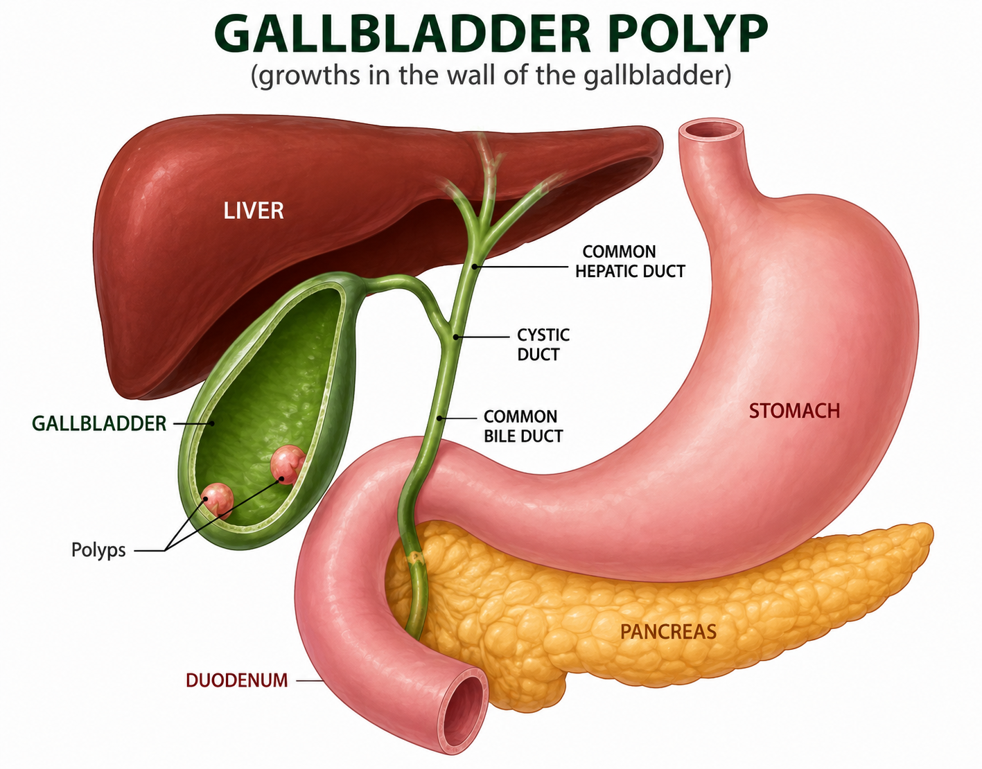
A gallbladder polyp originates from the gallbladder wall [2]. It projects from the lining into the cavity of the gallbladder and generally remains attached to the wall.
Both can be incidental findings, discovered during imaging performed for another medical reason. This does not mean that either should be ignored, but it also does not mean that every gallstone or polyp requires treatment. The significance of the finding depends on its characteristics, the patient's symptoms and the risk of future complications.
Bile contains cholesterol, bile salts, bilirubin and other substances. Gallstones develop when conditions within the bile favour crystallisation and the gradual formation of solid deposits. Cholesterol stones are the most common type in many populations, while pigment stones, which contain bilirubin-related material, form through different mechanisms.
Gallstones vary considerably in size and number. Some people have a single stone, while others develop multiple stones [3]. Importantly, many gallstones never cause symptoms and are discovered incidentally.
Several factors are associated with an increased likelihood of gallstone formation, including:
The presence of gallstones becomes particularly important when they interfere with the normal flow of bile. A stone obstructing the outlet of the gallbladder can produce biliary pain, while persistent obstruction can lead to inflammation of the gallbladder [4]. Stones that enter the bile ducts can cause further complications, including bile duct obstruction, infection or pancreatitis.
For this reason, doctors do not determine treatment simply by asking whether stones are present. Whether they are causing symptoms or complications is usually much more important.
Gallbladder polyps require a different type of assessment because they arise from the gallbladder wall instead of forming within bile. They appear on imaging as projections extending from the inner surface of the gallbladder into its cavity.
Importantly, most gallbladder polyps detected on ultrasound are benign [5]. Many are cholesterol polyps, sometimes called pseudopolyps, which are related to cholesterol deposits within the gallbladder wall. Other benign lesions include inflammatory polyps, while true neoplastic polyps such as adenomas are less common.
The reason polyps are assessed carefully is that some neoplastic lesions, including gallbladder cancer, can have a polypoid appearance. A diagnosis of a gallbladder polyp, therefore, does not mean that cancer is present. Instead, the purpose of assessment is to identify the relatively small group of lesions with features that warrant closer surveillance, further investigation or surgery.
Doctors may consider factors such as:
Most gallbladder polyps are asymptomatic and discovered incidentally. Depending on their characteristics and the patient's risk profile, management can range from no further follow-up to repeat ultrasound surveillance or consideration of gallbladder removal.
The distinction between gallstones and gallbladder polyps becomes particularly clear on ultrasound. Stones usually lie freely within the gallbladder and move with changes in body position, whereas polyps remain attached to the gallbladder wall. Their clinical significance is also assessed differently.
| Feature | Gallstones | Gallbladder Polyps |
| Origin | Form from the components of bile | Arise from the gallbladder wall |
| Position | Lie within the gallbladder cavity | Project from and remain attached to the gallbladder wall |
| Movement on Ultrasound | Usually move when the patient's position changes | Usually remain fixed to the wall |
| Acoustic Shadowing | Typically produce posterior acoustic shadowing | Typically do not produce posterior acoustic shadowing |
| Symptoms | Often asymptomatic but can cause biliary pain and complications | Usually asymptomatic |
| Main Clinical Concern | Symptoms and stone-related complications | Size, growth, morphology and features associated with malignancy risk |
| Monitoring | Routine surveillance is generally unnecessary for uncomplicated asymptomatic gallstones | Selected polyps may require follow-up ultrasound |
| Surgery | Usually considered when stones cause symptoms or complications | May be considered for polyps with characteristics that warrant removal |
Many gallstones remain silent, meaning they cause no symptoms and may never create a health problem. They are often discovered incidentally during an ultrasound or another abdominal investigation. The situation changes when a stone interferes with the normal drainage of bile.
A common presentation is biliary colic, which occurs when a gallstone temporarily obstructs the outlet of the gallbladder. This typically causes steady pain in the upper abdomen, often towards the right side or centre. The pain may become intense and can last from around 30 minutes to several hours before settling as the obstruction resolves.
More persistent obstruction or movement of stones into the bile ducts can lead to complications, including:
This distinction is important when considering treatment. Having gallstones is not the same as having symptomatic gallstone disease. An incidental stone that has never caused symptoms is approached differently from gallstones causing recurrent pain, inflammation or other complications.
With gallbladder polyps, doctors are usually less concerned with symptoms alone and more interested in what the lesion looks like and whether it has features associated with a greater likelihood of neoplasia or malignancy.
One of the most important characteristics is polyp size. Larger polyps warrant greater attention, and a size of around 10 mm has traditionally been an important threshold at which cholecystectomy may be considered or recommended in several clinical guidelines [10]. However, size should not be interpreted in isolation, and recommendations differ between guidelines, particularly for polyps below this threshold.
Assessment may also take into account:
This is why two patients with gallbladder polyps may receive different recommendations even when neither has symptoms. A small polyp with reassuring features may require little or no surveillance, while a larger, growing or otherwise concerning lesion may warrant closer investigation or consideration of gallbladder removal.
Abdominal ultrasound is usually the primary imaging test for suspected gallstones and gallbladder polyps. It allows the gallbladder, its contents and its wall to be examined without radiation and can often distinguish between the two findings.
Gallstones typically appear as bright, echogenic structures within the gallbladder. They usually move when the patient’s position changes and commonly produce posterior acoustic shadowing, where the stone blocks the ultrasound beam and creates a shadow behind it. Ultrasound can also identify findings that may accompany acute gallbladder inflammation, such as wall thickening or fluid around the gallbladder.
Gallbladder polyps behave differently on ultrasound. They:
Accurate measurement is particularly important for polyps because size can influence decisions about surveillance and surgery.
Sometimes an ultrasound cannot confidently determine the nature of a gallbladder lesion. In selected cases, additional or more specialised imaging may be considered to clarify an uncertain finding or provide further characterisation before management is decided.
Not every gallstone or gallbladder polyp requires treatment simply because it has been detected. The appropriate approach depends on the type of finding, whether it is causing symptoms, its imaging characteristics and the likelihood of future complications.
For gallstones, symptoms are an important part of the decision. Gallstones discovered incidentally in someone who has never experienced biliary symptoms can often be managed without surgery. Once stones begin causing recurrent pain or complications, however, treatment becomes more likely to be recommended.
Management of gallstones may therefore include:
For gallbladder polyps, management is guided less by symptoms alone and more by the characteristics of the lesion. The size and morphology of the polyp, changes seen on follow-up imaging and relevant patient-specific risk factors all help determine whether observation or surgery is appropriate.
Depending on these findings, management may involve:
The important distinction is that gallstones are generally treated because of the symptoms or complications they cause, while gallbladder polyps are managed primarily according to their characteristics and potential clinical risk. In both cases, the decision should be individualised instead of being based on the ultrasound finding alone.
Although gallstones and gallbladder polyps are different conditions, both may be treated with cholecystectomy when there is a clear clinical reason to remove the gallbladder.
Gallbladder removal may be recommended for:
Cholecystectomy removes the entire gallbladder together with the stones or polyp, instead of removing the individual finding alone. When surgery is appropriate, a laparoscopic approach is generally preferred where it can be performed safely.
An incidental gallstone or gallbladder polyp may cause no immediate problems. However, certain symptoms should not be ignored, particularly because they may indicate gallstone obstruction, inflammation or infection.
Seek prompt medical assessment if you develop:
These symptoms can occur with complications such as acute cholecystitis, bile duct obstruction, cholangitis or pancreatitis and may require urgent assessment. An incidental gallbladder polyp alone would not usually be expected to cause an acute episode of severe illness, so other biliary or abdominal causes need to be considered.
Gallstones and gallbladder polyps may both be identified during an ultrasound, but the similarity largely ends there. Gallstones form within bile and become clinically important mainly when they cause symptoms, obstruction or complications. Gallbladder polyps arise from the gallbladder wall and are assessed principally according to their size, morphology, growth and other features that help determine their level of risk.
Neither finding automatically means that surgery is necessary. Many people with asymptomatic gallstones require no treatment, while many small gallbladder polyps can be managed conservatively, depending on their characteristics and individual risk factors. An ultrasound result, therefore, needs to be considered alongside your symptoms, medical history, previous imaging and the specific features reported on the scan.
If an ultrasound has identified gallstones or a gallbladder polyp and you are unsure what the finding means for your health, consider scheduling a consultation with a specialist at Tan Siong San Surgery for an individual assessment and discussion of the appropriate next steps.


Tan Siong San Surgery. © All Rights Reserved 2025


