Liver Cysts: When They are Harmless and When They Need a Specialist’s Opinion
Home » Media » Liver Cysts: When They are Harmless and When They Need a Specialist’s Opinion
October 23, 2026
At a glance
Overview
Details
Condition
Liver cysts (hepatic cysts)
What They Are
Fluid-filled spaces that develop within the liver
How They Are Often Found
Incidentally during an ultrasound, CT scan or MRI performed for another reason
Are They Usually Harmful?
Most simple liver cysts are benign and cause no symptoms or complications
Do Simple Cysts Need Treatment?
Usually not if they have typical imaging features and are not causing symptoms
Is Routine Follow-Up Needed?
Routine imaging follow-up is generally not recommended for an asymptomatic, confidently diagnosed simple hepatic cyst
When Further Assessment May Be Needed
When the cyst causes symptoms, develops a complication or has atypical or complex imaging features
Possible Treatments for Symptomatic Simple Cysts
Depending on the individual case, options may include aspiration with sclerotherapy or surgical fenestration
When to Seek Medical Attention
Significant or worsening upper abdominal pain, fever, jaundice, sudden severe pain or other new concerning symptoms
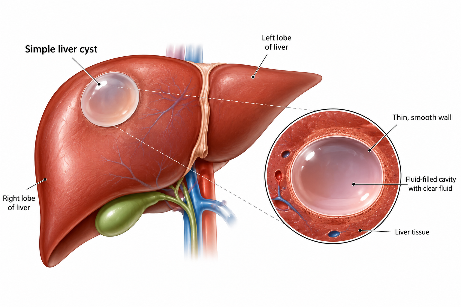
A simple liver cyst is a fluid-filled cavity within the liver that is usually benign and often discovered incidentally during abdominal imaging.
Liver cysts are frequently discovered incidentally during an ultrasound, CT scan or MRI performed for another reason. For many patients, the finding is unexpected and understandably raises questions about whether the cyst is dangerous, whether it could become cancerous and whether it needs to be removed.
In most cases, an incidental liver cyst is a simple hepatic cyst—a benign, fluid-filled lesion that causes no symptoms and does not require treatment. However, not every cystic lesion in the liver is a simple cyst, and the significance of the finding cannot be determined from the word “cyst” alone. Its appearance on imaging, the presence of symptoms and any associated complications are important in determining whether further assessment is necessary.
This is why management can differ considerably between patients. A typical simple cyst in someone without symptoms may require no further investigation or routine follow-up, whereas a symptomatic cyst or one with atypical imaging features may need additional imaging and specialist evaluation.
In this blog, we explain how simple liver cysts differ from other cystic liver lesions, when they can safely be left alone, which symptoms and imaging findings warrant closer attention and when a hepatobiliary specialist’s opinion may be appropriate.
What Exactly is a Liver Cyst?
A liver cyst, also called a hepatic cyst, is a fluid-filled cavity that develops within the liver. There are several types of cystic liver lesions, but the most common is the simple hepatic cyst, a benign cyst that is often discovered incidentally during imaging performed for another reason [1].
Simple hepatic cysts are believed to develop from small bile duct structures during development, although they do not usually communicate with the bile ducts themselves. A person may have a single cyst or several cysts, and most cause no symptoms or disruption to normal liver function.
Simple liver cysts also tend to have a recognisable appearance on imaging. On ultrasound, they typically contain clear fluid and appear as well-defined lesions with thin, smooth walls and no internal solid components or divisions. When these typical features are present in a person without symptoms, further investigation is often unnecessary.
Not all cystic lesions have this straightforward appearance, however. A cyst containing internal divisions, thickened or irregular walls, solid areas or other unusual features may represent a different type of liver lesion or a complicated cyst [2]. In these situations, further imaging with CT or MRI may be needed to establish the nature of the cyst and determine whether specialist assessment is appropriate.
Do Simple Liver Cysts Need Treatment?
In most cases, they do not. A simple liver cyst is a benign lesion, and when it has the characteristic appearance of a simple hepatic cyst on imaging and is not causing symptoms, treatment is generally unnecessary [3]. Most remain clinically insignificant and do not interfere with normal liver function or lead to complications.
Even if a simple cyst increases in size over time, this does not automatically mean that treatment is required. The decision is guided more by whether the cyst is causing symptoms, affecting nearby structures or developing complications than by a change in size alone. Similarly, once an asymptomatic cyst has been confidently identified as a simple hepatic cyst, routine imaging follow-up is generally not required.
Observation is therefore appropriate for most people with uncomplicated simple liver cysts. The approach may change if symptoms develop, complications such as bleeding or infection are suspected, or subsequent imaging raises uncertainty about the nature of the lesion. In these circumstances, further investigation or specialist assessment may be appropriate.
Distinguishing Simple from Complex Liver Cysts
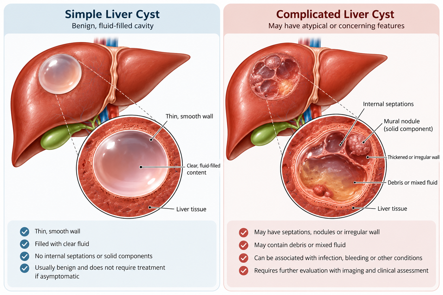
The characteristics of a liver cyst on imaging are central to determining its likely nature and whether further assessment is necessary. A typical simple hepatic cyst has a well-defined, thin and smooth wall, contains uniform fluid and has no internal septations or solid components. These features generally support a benign diagnosis when considered alongside the patient’s clinical presentation.
By contrast, findings such as internal septations, wall thickening or irregularity, mural nodules, calcification or internal debris may indicate a complicated cyst or another type of cystic liver lesion [4]. Such findings do not necessarily indicate malignancy, but they may warrant additional imaging or specialist assessment to establish the diagnosis and determine appropriate management.
Feature
Typical Simple Liver Cyst
Features That May Require Further Assessment
Contents
Clear, uniform fluid
Internal debris, blood or more complex contents
Wall
Thin and smooth
Thickened or irregular wall
Internal Divisions
Usually absent
Septations or multiple compartments
Solid Components
Absent
Mural nodules or solid components
Contrast Enhancement
No enhancement of the cyst contents
Enhancing walls, septa or solid tissue may require investigation
Symptoms
Usually none
Pain, pressure symptoms, fever or other associated symptoms may require assessment
Usual Approach
Observation if asymptomatic
Further characterisation and specialist assessment may be required
When atypical features are present, further evaluation may be needed to clarify the nature of the lesion and distinguish a complicated simple cyst from other cystic liver conditions. Depending on the initial findings, CT or MRI may be used to characterise the cyst in greater detail and guide subsequent management.
Simple liver cysts usually contain uniform fluid and have a thin, smooth wall, while septations, wall irregularity, nodules or other complex features may warrant further assessment.
Does the Size of a Liver Cyst Determine Whether It Is Dangerous?
It is natural to focus on size when a scan describes a liver cyst as “large”, but size alone does not determine whether a cyst is concerning or needs treatment. A large simple cyst may remain harmless and cause no symptoms, while a much smaller cyst may need closer assessment if it has unusual features on imaging.
Instead of looking at the measurement alone, doctors consider the cyst as a whole. Important questions include:
Does it have the typical appearance of a simple liver cyst?
Is it causing pain, abdominal fullness or other symptoms?
Is it pressing on the stomach, bile ducts or other nearby structures?
Are there signs of bleeding, infection or another complication?
Does it contain septations, nodules or other atypical features?
Is it a single cyst, or are there multiple cysts that may suggest another cystic liver condition?
Size becomes more relevant when a cyst begins to have a physical effect. A sufficiently large cyst may stretch the liver capsule or compress nearby structures, potentially causing pain, abdominal fullness, early satiety or, in some cases, problems with bile drainage. In these situations, treatment may be considered because of what the cyst is causing, rather than simply because it has reached a particular size.
When Can a Liver Cyst Start Causing Symptoms?
Most simple liver cysts cause no symptoms at all. When symptoms do occur, they are more likely to arise when a cyst becomes sufficiently large to exert pressure on surrounding structures or when a complication develops [5].
Possible symptoms include:
Dull or persistent discomfort in the right upper abdomen
A sensation of abdominal fullness or pressure
Abdominal distension
Feeling full sooner than expected when eating
Nausea in some patients
These symptoms are not specific to liver cysts. Gallbladder disease, stomach conditions, bowel disorders and many other abdominal problems can produce similar complaints. Finding a cyst on a scan therefore does not prove that the cyst is responsible for a patient’s symptoms.
This is particularly important when treatment is being considered. A specialist assessment can help determine whether the location and size of the cyst plausibly explain the symptoms or whether another cause needs to be investigated.
What Complications Can Develop from a Liver Cyst?
Most simple liver cysts remain uncomplicated and never cause significant problems. Complications are uncommon, but they can occur, particularly in larger or symptomatic cysts. When they do, the symptoms and appearance of the cyst may change and further medical assessment may be necessary.
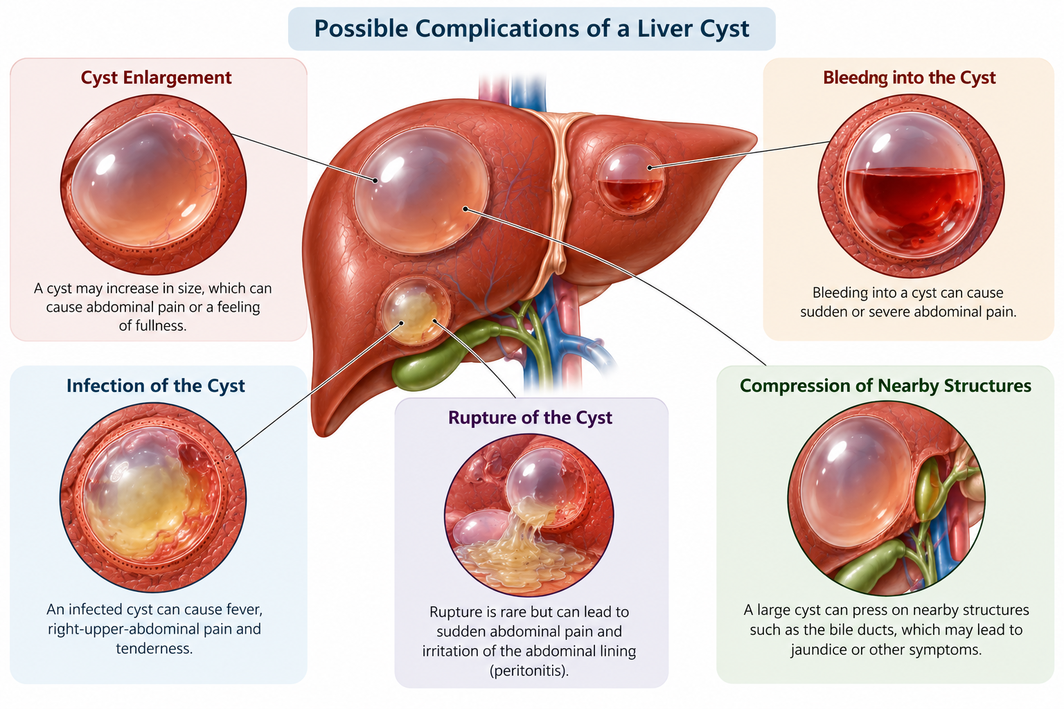
Bleeding into the cyst — Occasionally, bleeding can occur within a liver cyst, known as intracystic haemorrhage [6]. This may cause sudden or worsening upper abdominal pain and can change the internal appearance of the cyst on imaging. Because bleeding can make a previously simple cyst appear more complex, additional imaging may be needed to confirm the cause of the change and exclude other cystic lesions.
Cyst infection — A liver cyst can become infected, although this is uncommon. Infection may cause fever, upper-right abdominal pain or tenderness and other signs of inflammation or infection [7]. Changes may also be visible within or around the cyst on imaging. An infected cyst requires medical assessment and appropriate treatment rather than continued observation alone.
Compression of nearby structures — A large cyst may place pressure on surrounding organs or structures. Depending on its size and location, this can contribute to persistent abdominal discomfort, fullness or early satiety. Less commonly, compression of the bile ducts can interfere with bile drainage and contribute to jaundice.
Cyst rupture — Rarely, a liver cyst can rupture, allowing its contents to leak into the abdominal cavity [8]. This may cause sudden abdominal pain and requires prompt medical assessment, particularly when the pain is severe or accompanied by other new symptoms.
The development of a complication changes how a liver cyst is managed. A cyst that was previously suitable for observation may require further imaging, specialist assessment or treatment if bleeding, infection, rupture or significant compression develops.
Large or complicated liver cysts can occasionally produce pain or pressure by stretching the liver capsule or compressing nearby abdominal structures.
When Does a Liver Cyst Need a Specialist’s Opinion?
Not every incidental simple cyst needs specialist referral simply because it has been identified. A specialist opinion becomes more relevant when there is uncertainty about what the lesion is, whether it is responsible for symptoms or whether intervention may be necessary.
Further assessment may be appropriate when:
The cyst has septations, wall thickening, irregularity, nodules or other complex features
The diagnosis of a simple cyst is uncertain
Persistent or recurrent symptoms may be attributable to the cyst
A large cyst is compressing nearby structures
There is concern about bleeding into the cyst
Infection is suspected
Jaundice or evidence of bile duct obstruction develops
There are numerous liver cysts suggesting polycystic liver disease
Imaging raises the possibility of a parasitic or cystic neoplastic lesion
Treatment of a symptomatic cyst is being considered
The purpose of specialist assessment is not necessarily to recommend surgery. In many cases, it is to establish what type of cyst is present and whether anything needs to be done at all.
A hepatobiliary surgeon can review the imaging alongside the patient’s symptoms, medical history and other investigations to determine whether observation, additional imaging or treatment is appropriate.
Not Every Cystic Liver Lesion is a Simple Cyst
Although simple hepatic cysts are benign, not every fluid-containing or cyst-like lesion identified in the liver belongs to this category. Several other conditions can have a cystic appearance on imaging, which is why lesions with atypical features may require further investigation before their nature can be established.
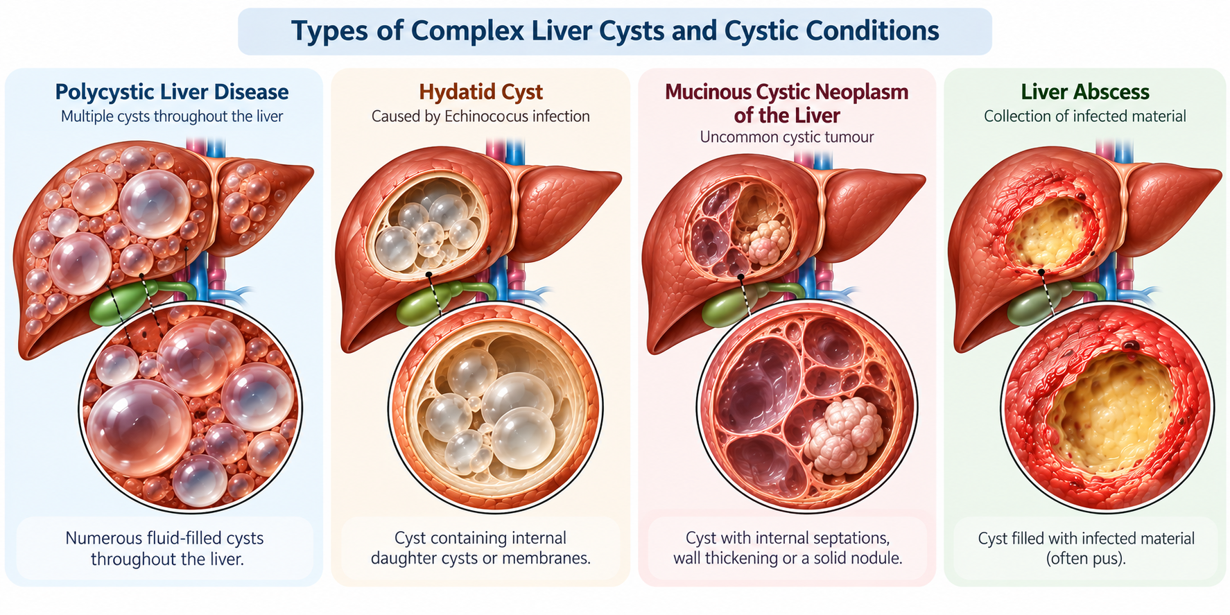
Polycystic liver disease — Unlike an isolated simple cyst, polycystic liver disease is characterised by the presence of numerous cysts throughout the liver [9]. It can occur as a separate inherited condition or in association with autosomal dominant polycystic kidney disease. Liver function is often preserved, although extensive cysts can enlarge the liver and cause abdominal fullness, discomfort or other pressure-related symptoms.
Hydatid cysts — These cysts develop as a result of infection with Echinococcus parasites and are managed differently from simple hepatic cysts [10]. A person’s exposure or travel history, together with characteristic findings on imaging, can help doctors determine whether hydatid disease needs to be considered.
Mucinous cystic neoplasms of the liver — These are uncommon cystic tumours that require distinction from simple liver cysts because their management is different [11]. Imaging may reveal features such as internal septations, wall thickening or mural nodules. When these or other suspicious features are present, further characterisation is important before treatment is planned.
Liver abscesses — A liver abscess is a collection of infected material within the liver rather than a true simple cyst. It may be associated with fever, abdominal pain, tenderness and other signs of infection. Imaging, together with the patient’s symptoms and clinical findings, helps distinguish an abscess from other cystic liver lesions.
These conditions do not mean that an incidental liver cyst should automatically be considered concerning. Rather, they highlight the importance of establishing whether a lesion has the typical features of a simple hepatic cyst.
When the appearance is atypical or the diagnosis remains uncertain, further imaging or specialist assessment may be needed to determine the nature of the lesion and the appropriate next step.
Complex liver cystic conditions include polycystic liver disease, hydatid cysts, mucinous cystic neoplasms and liver abscesses, each of which has distinct causes, imaging features and management considerations.
Do You Need Regular Scans for a Simple Liver Cyst?
For most people with an asymptomatic simple liver cyst, routine follow-up scans are not necessary. Once imaging has clearly established that the lesion has the typical features of a simple hepatic cyst, and there are no associated symptoms or complications, regular ultrasound, CT or MRI solely to monitor the cyst is generally not recommended.
This is because simple cysts are benign, and changes in size alone do not usually determine whether treatment is required. Repeatedly measuring a cyst that continues to have a typical appearance and causes no symptoms is therefore unlikely to change its management. Instead, further imaging is usually guided by a change in the clinical situation.
Repeat imaging may be appropriate if:
New or persistent symptoms develop
The initial scan does not clearly establish that the lesion is a simple cyst
Atypical or complex features require further characterisation
Bleeding, infection or another complication is suspected
Another liver condition requires ongoing imaging surveillance
A specialist recommends reassessment based on the individual clinical findings
The need for follow-up therefore depends on why another scan is being performed. Routine surveillance is generally unnecessary for a confirmed, asymptomatic simple hepatic cyst, whereas new symptoms, uncertain imaging findings or suspected complications may provide a clear reason for further investigation.
How are Liver Cysts Diagnosed and Assessed?
Liver cysts are usually identified through an abdominal ultrasound, although they may also be detected on CT or MRI performed for another reason. Once a cyst is found, the main purpose of assessment is to determine whether it has the typical features of a simple hepatic cyst or requires further investigation.
Doctors may consider:
The appearance of the cyst — A simple cyst typically has a thin, smooth wall, uniform fluid contents and no internal septations or solid components.
Whether complex features are present — Septations, wall thickening or irregularity, mural nodules, debris or other atypical findings may require closer evaluation.
Whether symptoms are present — Persistent abdominal pain, fullness or other symptoms may influence whether further assessment is needed.
Whether complications are suspected — Findings suggesting bleeding, infection, rupture or compression of nearby structures may require additional investigation.
Whether further imaging is necessary — CT or MRI may be recommended when ultrasound does not clearly establish the nature of the cyst or when more detailed characterisation is required.
The wider clinical context — Medical history, previous imaging and relevant exposure or travel history may help distinguish a simple cyst from other cystic liver conditions.
For a clearly defined, asymptomatic simple hepatic cyst, ultrasound may be all that is required. Further investigation is generally reserved for cysts that are symptomatic, complicated or cannot be confidently characterised as simple.
What Treatment Options are Available for a Symptomatic Liver Cyst?
Most simple liver cysts do not require treatment. When a simple hepatic cyst is causing clinically significant symptoms or complications, however, treatment may be considered to reduce the cyst and relieve the problems associated with it.
The choice of treatment depends on the cyst’s size, location and anatomy, as well as the patient’s symptoms and overall clinical circumstances.
Aspiration with sclerotherapy — This minimally invasive approach involves draining fluid from the cyst using a needle or catheter placed under imaging guidance [12]. A sclerosing agent may then be introduced to act on the cyst lining and reduce the likelihood of fluid accumulating again. Aspiration alone is generally less effective as a long-term treatment because the cyst may refill when its fluid-producing lining remains intact.
Laparoscopic fenestration or deroofing — This procedure involves removing part of the cyst wall so that the cyst no longer remains a closed, fluid-filled cavity [13]. Where the location and anatomy of the cyst are suitable, fenestration may be performed using a laparoscopic, or keyhole, approach.
Liver resection — Removal of part of the liver is not routinely required for an uncomplicated simple hepatic cyst [14]. It may be considered in selected cases where the location or nature of the lesion makes other approaches unsuitable, or where the underlying diagnosis requires complete surgical removal.
The appropriate treatment is therefore individualised. A hepatobiliary specialist will consider the type of cyst, the symptoms or complications it is causing, its location and anatomy, and the relative benefits and risks of the available treatment options before recommending an approach.
Key Takeaway
Most liver cysts are simple hepatic cysts that are benign, cause no symptoms and require no treatment. When a cyst has the typical features of a simple cyst on imaging and there are no associated symptoms or complications, routine follow-up scans are generally unnecessary. Size alone is also not an indication for treatment; even a relatively large simple cyst may be managed conservatively if it is not causing problems.
Further assessment becomes important when the clinical or imaging picture is less straightforward. Persistent symptoms, compression of nearby structures, bleeding, infection or rupture may change how a previously simple cyst is managed.
Similarly, features such as internal septations, wall thickening or irregularity, nodules or solid components may require additional imaging to distinguish a complicated simple cyst from other cystic liver conditions. When treatment is necessary for a symptomatic simple cyst, options such as aspiration with sclerotherapy or laparoscopic fenestration may be considered depending on the individual case.
If you have been diagnosed with a liver cyst and are unsure what the finding means or whether further assessment is necessary, consider scheduling a consultation with a hepatobiliary specialist at Tan Siong San Surgery for an individual assessment of your imaging findings and advice on whether observation, further investigation or treatment may be appropriate.
Frequently Asked Questions (FAQs)
Can a simple liver cyst turn into cancer?
A true simple hepatic cyst is benign and is not regarded as a precancerous lesion. The important issue is ensuring that a cystic lesion with complex or suspicious features has not been incorrectly classified as a simple cyst. If the imaging appearance is atypical, further characterisation may therefore be recommended.
Does a large liver cyst always need to be removed?
No. Size alone does not automatically determine whether treatment is necessary. A large cyst that has typical simple features and causes no symptoms may not require intervention. Treatment becomes more relevant when a cyst produces symptoms, compresses surrounding structures, develops a complication or has uncertain characteristics.
Can a liver cyst cause abnormal liver function tests?
Most uncomplicated simple hepatic cysts do not interfere with normal liver function. Abnormal liver tests should therefore not automatically be attributed to a cyst. In unusual cases, a large or strategically located cyst can compress the biliary system, but other causes of abnormal liver tests also need to be considered.
Can liver cysts disappear on their own?
Simple hepatic cysts generally persist once they have formed, although their size may remain stable or change over time. Their continued presence does not itself mean that treatment is necessary.
Can liver cysts grow?
Yes. Some simple hepatic cysts can enlarge over time. Growth does not automatically indicate cancer or create a need for surgery. Symptoms and imaging characteristics are more important in determining whether further assessment or treatment is appropriate.
Can a liver cyst cause pain?
It can, particularly if the cyst becomes large enough to stretch the liver capsule or compress nearby structures. Bleeding, infection or rupture can also cause pain. However, abdominal pain has many possible causes, so the presence of a liver cyst does not necessarily prove that the cyst is responsible.
Is an ultrasound enough to diagnose a liver cyst?
Ultrasound is generally the first-line imaging test and can be sufficient when a simple hepatic cyst has characteristic uncomplicated features. CT or MRI may be appropriate when symptoms are present, complications are suspected or ultrasound shows atypical features requiring further characterisation.
Do simple liver cysts need yearly ultrasound scans?
Routine imaging follow-up is generally not recommended for an asymptomatic simple hepatic cyst once the diagnosis is secure. Further imaging may be performed if symptoms develop or if there is another clinical reason to reassess the lesion.
Is draining a liver cyst enough to treat it?
Simply aspirating the fluid may not provide lasting treatment because the cyst can refill. When percutaneous treatment is appropriate, aspiration may therefore be combined with sclerotherapy. Surgical fenestration is another treatment option for selected symptomatic simple cysts.
What kind of doctor should assess a complicated liver cyst?
A hepatobiliary specialist may be appropriate when a cyst is symptomatic, has complex imaging characteristics, causes complications or may require intervention. Hepato-pancreato-biliary (HPB) surgeons specialise in conditions affecting the liver, gallbladder, bile ducts and pancreas.
References
Rawla, P., Sunkara, T., Muralidharan, P., & Raj, J. P. (2019). An updated review of cystic hepatic lesions. Clinical and Experimental Hepatology, 5(1), 22–29. https://doi.org/10.5114/ceh.2019.83153
Baltagiannis, E. G., Tsili, A., Goussia, A., Glantzouni, A., Frigkas, K., Charchanti, A., Glantzounis, G. K., & Gomatos, I. P. (2024). Complex cystic liver lesions: Classification, diagnosis, and management. Annals of Gastroenterology, 37(3), 255–265. https://doi.org/10.20524/aog.2024.0876
Rho, S. Y., Lee, J.-M., Park, M.-S., Kang, W.-H., Byun, Y., Lee, S. J., Shin, A., Yoon, J. H., Kim, B., & Yi, N.-J. (2025). Diagnosis, treatment and prognosis of simple hepatic cyst: Clinical practice guideline. Annals of Hepato-Biliary-Pancreatic Surgery, 29(3), 218–225. https://doi.org/10.14701/ahbps.25-070
Loaiza, S. C., Torre, C. de la, Lozada, L. R., & Moslero, M. C. C. (2014, March 6). Differential diagnosis of complex liver cysts. European Congress of Radiology - ECR 2014. ECR 2014 EPOS. https://epos.myesr.org/poster/esr/ecr2014/C-0801
Agrawal, A., Elnashar, M., Garg, K., Mustafa, A., Rosenzveig, A., Arockiam, A. D., Haroun, E., Khurana, R., Klein, A. L., & Wang, T. K. M. (2026). Contemporary review of clinical features, multi-modality imaging, and management of pericardial cysts. Journal of Clinical Medicine, 15(4), 1585. https://doi.org/10.3390/jcm15041585
Purdy, A. C., Grigorian, A., Fernando, D., Nahmias, J., & Demirjian, A. N. (2019). Intracystic hemorrhage complicating polycystic liver disease in a 90-year-old woman. ACG Case Reports Journal, 6(4), e00049. https://doi.org/10.14309/crj.0000000000000049
Miliadis, L., Giannakopoulos, T., Boutsikos, G., Terzis, I., & Kyriazanos, I. D. (2010). Spontaneous rupture of a large non-parasitic liver cyst: A case report. Journal of Medical Case Reports, 4, 2. https://doi.org/10.1186/1752-1947-4-2
Norcia, L. F., Watanabe, E. M., Hamamoto Filho, P. T., Hasimoto, C. N., Pelafsky, L., de Oliveira, W. K., & Sassaki, L. Y. (2022). Polycystic liver disease: Pathophysiology, diagnosis and treatment. Hepatic Medicine : Evidence and Research, 14, 135–161. https://doi.org/10.2147/HMER.S377530
Xu, Y., Hu, X., Li, J., & Dong, R. (2020). Hepatic hydatid cyst misdiagnosed as simple cyst: A case report. The American Journal of Case Reports, 21, e923281-1-e923281-6. https://doi.org/10.12659/AJCR.923281
Hutchens, J. A., Lopez, K. J., & Ceppa, E. P. (2023). Mucinous cystic neoplasms of the liver: Epidemiology, diagnosis, and management. Hepatic Medicine : Evidence and Research, 15, 33–41. https://doi.org/10.2147/HMER.S284842
Dong, Z., Zhao, M., Chen, J., Zhao, Y., Sun, X., Huang, B., & He, Y. (2025). Giant cysts, no incisions: Ultrasound-guided sclerotherapy in the elderly. Medicine, 104(52), e46897. https://doi.org/10.1097/MD.0000000000046897
Bernts, L. H. P., Echternach, S. G., Kievit, W., Rosman, C., & Drenth, J. P. H. (2019). Clinical response after laparoscopic fenestration of symptomatic hepatic cysts: A systematic review and meta-analysis. Surgical Endoscopy, 33(3), 691–704. https://doi.org/10.1007/s00464-018-6490-8
Macedo, F. I. (2013). Current management of noninfectious hepatic cystic lesions: A review of the literature. World Journal of Hepatology, 5(9), 462–469. https://doi.org/10.4254/wjh.v5.i9.462
This article has been medically reviewed by Dr. Tan Siong San
Dr Tan is a Senior Consultant Surgeon with over 25 years of clinical experience in the field of Hepato-Pancreato-Biliary (HPB) surgery treating conditions affecting gallbladder, liver and pancreas.